
Affiliations
ABSTRACT
Branchial cleft anomalies are uncommon in adults but common in children. Bilateral presentations are very rare especially bilateral cleft fistulae and sinuses simultaneously and mostly are a component of familial syndromes like branchio-oto-renal syndrome (BORS). We present a case of a 20-year male with no known comorbids complaining of painful swelling on the right side of neck along with draining fistula on the left side of neck. After a thorough history, neck examination, CT and x-ray fistulography, a final diagnosis of bilateral second branchial cleft fistulae and sinuses was made. The patient was put on operative list and after completing the preoperative workup, the surgical excision by step-ladder approach was done. Patient was discharged on 4th postoperative day after counselling him about the wound care. He was called for a follow-up after 7 days. On follow-up, the wound had started healing.
Key Words: Branchial fistula, Sinus, 2nd branchail arch, Bilateral.
INTRODUCTION
The branchial arch malformations are infrequent anomalies in the head and neck. Among branchial arch anomalies, second branchial arch anomalies are by far the most prevalent constituting 80-95% of the cases. Only 2 to 3% cases are bilateral. The branchial arches give rise to the face, neck, and the pharynx during the embryonic period.1 The anomalies develop as a result of incomplete involution of pharyngeal apparatus during the development.1,2 These anomalies include brachial cysts, fistulas, and sinuses. Fistulas, are seen in 7.5% of cases of bilateral second-branchial arch abnormalities.3 Sinuses are more frequent than cysts, which are more frequent than fistulae. Complete branchial fistula having external as well as internal openings is very rare.4,5
Branchial fistulae are typically diagnosed in infancy or childhood with outflow of purulent or mucoid discharge from a skin opening at the intersection of lower and middle third of sternocleidomastoid muscle at its anterior border.1,3 The detection of branchial cleft anomalies, especially branchial cleft fistula is very significant as it may be a component of a more extensive syndromic clinical presentation like branchio-oto-renal syndrome (BORS),2 which requires further investigations.
Since second branchial arch anomalies increase the risk of recurrent infections, the treatment of choice is complete surgical excision.3
We present a case of a 20-year male with painful swelling on the right side of neck along with draining fistula on the left side of neck. After necessary investigations, a final diagnosis of bilateral second branchial cleft fistulae and sinuses was made and managed successfully.
CASE REPORT
A 20-year male with no known comorbids presented with complaints of painful swelling on the right side of neck along with draining fistula on the left side of the neck. These swellings were present since his birth and were first noticed by his mother. The swelling on the right side gradually increased in size and became progressively painful, and after some time, it burst. The pain was not associated with coughing, eating, or swallowing. The swelling had a yellow-coloured, thick, foul-smelling discharge that was not associated with any activity. It was not associated with any aggravating or relieving factor. There was no swelling like this on any other part of the body. On the left side, the discharge was clear and non-foul smelling. The patient denied any family history. He was a non-smoker and had no history of addictions.
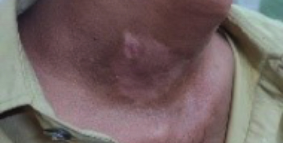
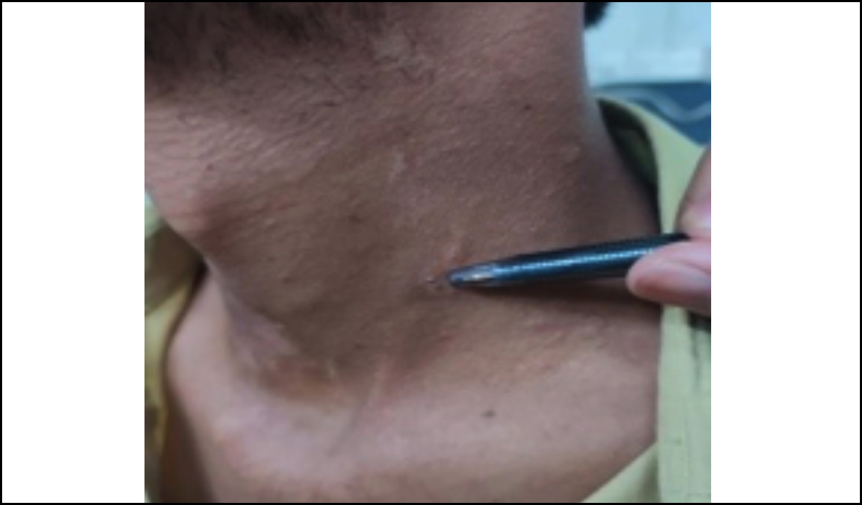
On neck examination, two discolourations on each side of the neck at the level of C6 and C7 vertebrae were noted. On the left side, it was located laterally at the level of cricoid cartilage. Another plaque was noted on the right side of neck at the same level. A 2×2 cm swelling was also observed on the right side of the neck with opening on the skin surface (Figure 1). No swelling was observed on the left side; only one discharging punctum was noted (Figure 2). Oral and nasal examinations were unremarkable. On ear examination, prominent malleus handle was present bilaterally, and whitish patch on tympanic membrane was present on the left side.
 Figure 1: Swelling and discolouration on the lateral aspect of the right side of neck.
 Figure 2: No swelling; only a punctum is seen on the left side of the neck.
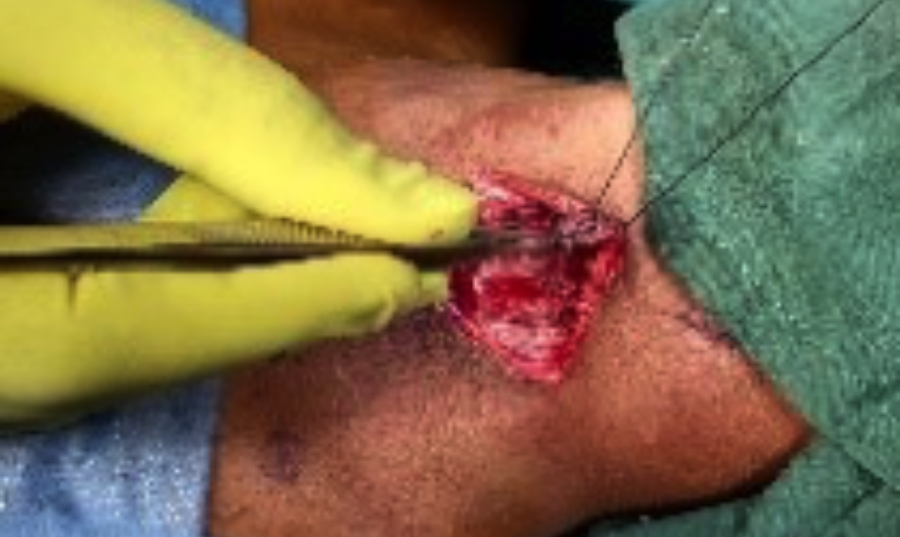
 Figure 3: Elliptical incision on the right side of neck along with tract identification.
Ultrasound of neck swelling showed a well-defined encapsulated hypoechoic lesion in the subcutaneous fat plane measuring 4.31 × 1.32 cm showing posterior acoustic shadowing. CT scan of neck with contrast showed a well-defined unilocular hypodense collection with thin enhancing wall measuring 1.5 × 4.6 × 6.2 cm in its anteroposterior, transverse and craniocaudal dimensions on the right side of the neck at the level of thyroid. Anteriorly, it was producing focal bulge on the overlying skin. Posteriorly, it was in close contact with the sternocleidomastoid (SCM) muscle. x-ray fistulogram showed an external opening on the left side of the neck at C6/7 level. A narrow regular tract was seen in soft tissues of anterolateral aspect of neck. It extended upward and slightly posterior to SCM muscle which opened into the oropharynx. The findings were suggestive of brachial fistula. On x-ray sinugram, external opening on the right anterolateral aspect of lower neck was identified and contrast medium was injected. An oblong collection measuring 3.4 × 1.9 × 1.3 cm was seen in soft tissues of neck on the right side, caudally extending down to supraclavicular region.
According to the above findings, the final diagnosis of brachial cleft fistula on the left and branchial cleft sinus on the right side was made. The patient was put on operative list, and preoperative workup was completed.
The operation was performed under general anaesthesia with orotracheal intubation. An elliptical incision was given on the left side of the neck around the opening of the swelling and fistula tract was identified and traced upwards (Figure 3). Another incision was given 5 cm above the previous incision to trace the tract. It was extended till the floor of mouth, and the whole tract of fistula was excised from the left side of the neck. On the right side, an elliptical incision was given, and the sinus was removed. 3/0 vicryl and 4/0 prolene sutures were used to close subcutaneous tissues and skin, respectively.
Histopathology showed left brachial fistula tract with features of lymphoepithelial cyst. It was negative for malignancy. Right-sided lesion showed infected fascia, and skin covered tissue showed acute and chronic inflammation, granulation tissue formation and sheets of foamy macrophages. Postoperatively, the patient was prescribed injection, Augmentin 1.2 g TDS for 7 days and injection paracetamol 1 g as per need. The patient was discharged on 4th postoperative day, was taught wound care, and called for follow-up after 7 days. On follow-up, the wound had started healing.
DISCUSSION
The formation of branchial arches, clefts, and pouches matches the development of gills in fish and amphibians but true gills (branchia) never develop in the human embryo. The branchial apparatus is comprised of six pairs of mesodermal arches that are bordered inside by endodermal pouches and externally by ectodermal clefts. The formation of the branchial apparatus initiates during the second week of embryonic development and is finished by the sixth to seventh week.1,4
In rare cases, branchial cleft abnormalities, especially bilateral branchial cleft fistulae, may be a component of a syndrome like - BORS characterised by an association of: 1) branchial fistulae or cysts or sinuses; 2) ear abnormalities, which can include the inner, middle and outer ear; and 3) renal malformations, which can range in severity from renal hypoplasia to agenesis.2 BORS syndrome is an autosomal dominant disorder. Mutations in the human homologue of the Drosophila eyes absent gene (EYA1) are the most common cause of BORS.6 This patient presented with left-sided branchial cleft fistula and right-sided branchial sinus and he did not exhibit any other features of BORS. Also, there was no family history of such findings. The current case showed the existence of bilateral branchial cleft sinus and fistula simultaneously making it a rare scenario.
Branchial cleft fistula can be diagnosed on the basis of clinical history and examination. However, various imaging modalities can be used such as fistulography, CT scan/MRI in order to define the extent of the lesion and confirm the diagnosis. But, literature regarding the pre-operative tools for diagnostic accuracy varies. In a study by Mitroi et al., the authors demonstrated 100% accuracy of ultrasound in the identification of 23 branchial cysts and sinuses, eliminating the requirement for a CT or MRI scan.7 Other authors reported a significantly lower accuracy stating operator dependency as a prime cause. Regarding CT scan, in a retrospective study by Schroeder et al., in which 87 patients with branchial cleft anomalies were reviewed, the diagnostic accuracy for cysts was 95%, for sinuses was 81%, and for fistulae, it was 50% on preoperative CT scans.8 MRI can accurately identify branchial cysts; however, its efficacy in diagnosing fistulae and sinus tracts can vary.9 Regarding fistulography with x-ray and the use of intraoperative fistulograms, which employ plain film radiography and water-soluble contrast, just before the surgical excision, had been suggested by authors as being well tolerated, quick, and affordable.10
For all branchial anomalies, surgical excision is the only effective treatment as they do not regress spontaneously. Among the various approaches, the most frequent one is via an elliptical incision made at the level of tract’s external opening as was done in this case. Historically, sclerosing agents were injected in the fistulae in order to treat with minimal scarring but a potent reaction was produced, and the approach was abandoned since there was a possibility that significant adjacent structures may sustain damage or pharyngeal perforation can occur.
PATIENT’S CONSENT:
Written, informed consent was taken from parents of the patient to publish this case report.
COMPETING INTEREST:
The authors declared no conflict of interest.
AUTHORS’ CONTRIBUTION:
SQ: Manuscript writing.
SI: Discussion writing.
AH: Literature search.
HA: Final approval
All authors approved the final version of the manuscript to be published.
REFERENCES